If you’ve ever had a root canal treatment, or noticed recognizing early root canal symptoms
before seeking care, you might have envied the precision displayed during treatment. At Smile Craft Advanced Dental Clinic, we offer specialized laser and microscope-assisted root canal procedures to add maximum accuracy to endodontic care.. This high-tech tool helps dentists achieve exceptional precision, ensuring that your dental experience is as smooth and effective as possible. With the help of magnification and illumination, dentists can see the tiniest details within the tooth, making a remarkable difference in outcomes. In this blog, we’ll explore how this dental technology works and why it brings a new level of precision to root canal treatments.
Understanding Microscope-Assisted RCT
Image courtesy: Unsplash
Definition and Purpose of Microscope-Assisted RCT
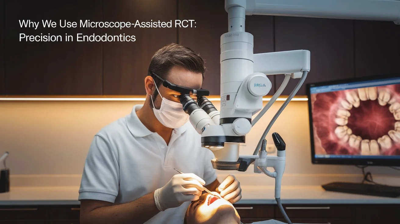
Alright, let’s start with the basics! Microscope-Assisted Root Canal Treatment (RCT) is like giving dental professionals a superpower. Imagine having a tool that transforms your dentist into a miniaturized action hero, diving deep into the tiny nooks and crannies of your tooth. That’s precisely what a microscope does in endodontics! This magical tool, equipped with high magnification and superior lighting, is used during root canal treatments to enhance dentists’ vision. The primary purpose? To ensure extreme precision during procedures, uncovering the finest details that the naked eye could easily miss. This meticulous approach helps in removing infected tissues, minimizing possible complications, and ensuring the tooth’s longevity.
Traditional RCT vs. Microscope-Assisted RCT
In the world of dentistry, not all root canals are created equal. When comparing the standard endodontic treatment process and pricing with high-precision methods, vision makes all the difference. While conventional approaches clear infections, incorporating principles of modern minimally invasive dentistry ensures hidden complexities are never overlooked.
Enter Microscope-Assisted RCT—a game-changer in the field! Unlike the traditional approach, microscope-assisted RCT provides dentists with a detailed, illuminated view. It allows for a more comprehensive cleaning process, reaching into intricate spaces within the canal that might be overlooked otherwise. Think of it as swapping your regular spectacles for high-definition binoculars. This not only streamlines the procedure but also improves outcomes dramatically!
Technological Advances in Endodontics
The realm of dental technology is constantly evolving—and endodontics hasn’t been left behind. Alongside advanced laser therapy for endodontic care, the inclusion of operating microscopes in root canal therapy brings unprecedented vision. Features like variable magnifications, high-quality optics, and digital imaging give practitioners a clearer view inside complex root structures.
These technological marvels allow for a better understanding of complex root canal systems, enhancing the precision and success of the treatments.
Moreover, as we progress, other technologies like digital radiography and 3D imaging further boost diagnostic capabilities and treatment planning. Together with microscopes, they form a robust framework for endodontic excellence, leading to healthier smiles all around!
Benefits of Microscope-Assisted RCT
Enhanced Precision and Accuracy in Treatments
Imagine trying to solve a maze in the dark—it’s hard, right? Now imagine doing the same but with a flashlight—the experience is entirely different and far more successful. That’s what microscope-assisted RCT does for dentists. By illuminating and magnifying the root canals, dentists can thoroughly locate and remove infected tissues, avoiding the severe dangers of leaving severe dental infections untreated
. This meticulous inspection resolves deep-seated bacterial pockets, relieving issues like severe tooth pain that spikes at night and minimizing complications. In dentistry, precision is king, and microscope-assisted RCT delivers with royal aplomb.
Improved Visualization for Difficult Cases
Not all dental cases are straightforward. Sometimes, dentists face challenges such as calcified canals, unusual anatomies, or broken-off instruments. These scenarios demand more than just skill—they require enhanced visualization. Microscope-assisted RCT provides just that. With superior lighting and magnification, even the trickiest of cases can be addressed with confidence and clarity. It’s like turning on the lights when the path seems obscured.
This improved visualization translates to tackling complex situations efficiently, reducing the challenges associated with unforeseen surprises that the dentist might encounter during a procedure. And let’s not forget, when your dentist has such a keen view, patients can rest assured that they are receiving top-notch care!
Increased Success Rates and Patient Satisfaction
Ultimately, it’s all about the smiles—both the dentist’s and the patient’s. Microscope-assisted RCT raises the success bar by decreasing the chances of missing something important. This precision leads to fewer follow-up treatments and less post-procedure discomfort, ensuring that patients leave the office happier and healthier.
Moreover, knowing that their treatment involves cutting-edge technology gives patients peace of mind and boosts their trust in the process. When patients feel comfortable and confident with their care, their satisfaction naturally increases. After all, a content patient often turns into a loyal friend of the practice, ensuring that their next visit is more about routine check-ups than emergency treatments!
The Process of Microscope-Assisted RCT
Image courtesy: Unsplash

When most people think of root canal treatment (RCT), they probably envision a standard dental procedure. But did you know that with advancements in dental technology, this process has been revolutionized? Enter microscope-assisted RCT, a cutting-edge approach that elevates precision and outcomes.
Steps Involved in the Procedure
Embarking on a microscope-assisted RCT involves several meticulously executed steps:
- – Initial Examination: The dentist performs a thorough examination, often using digital imaging to assess the infected tooth and surrounding area.
- – Microscope Setup: The dental professional positions the microscope, which offers magnification and illumination of the tooth’s intricate canals—something a naked eye cannot achieve.
- – Access Opening & Cleaning: With precise tools and enhanced vision, the dentist creates a small opening to access the pulp chamber. Then comes cleaning, which involves removing infected tissue and bacteria from the root canals.
- – Shaping and Filling: The canals are shaped for optimal filling, ensuring no voids are left behind. Using microscopic detail, the root canals are then filled with a suitable material to seal them.
- – Restoration: Finally, the tooth structure is rebuilt. Depending on the remaining tooth structure, your dentist may place natural-looking tooth-coloured fillings
or fit protective dental crowns. If you are unsure which option suits your condition, choosing between a crown and a filling depends on the extent of structural reinforcement required.
Role of Dental Professionals and Training
Dental professionals play a pivotal role in ensuring the success of microscope-assisted RCT. Specialized training is essential as it equips them with the skills needed to harness the power of dental microscopes. This training covers the technical aspects of handling microscopes and the ability to make delicate decisions with unparalleled accuracy.
Courses and workshops offer hands-on experience, allowing practitioners to refine their skills and stay updated with the latest advancements in dental technology. This meticulous training ensures that patients receive the most precise care possible.
Expectations for Patients Undergoing the Treatment
Patients coming in for a microscope-assisted RCT can expect a smoother and more comfortable experience than traditional methods. Thanks to advanced imaging and microscope technology, dental professionals can complete procedures with increased accuracy, reducing the risk of complications.
Patients are often surprised by how quickly the procedure can be accomplished, and many report less post-operative discomfort. The precision of the treatment reduces the likelihood of recurring infections, promoting long-term dental health. After the procedure, patients can expect a brief recovery period and will usually be advised on how to care for their treated tooth to maximize its lifespan. With such sophisticated care, it’s no wonder patients leave feeling more confident with their dental health in check!
Conclusion
In summary, microscope-assisted root canal treatment (RCT) is a game-changer in the field of endodontics. By offering enhanced precision, it ensures that the intricacies of dental treatment are handled with the utmost care and effectiveness. With better diagnostics and treatment outcomes, patients enjoy healthier, longer-lasting results. As dental technology continues to advance, the importance of incorporating tools like microscopes into everyday practice cannot be overstated. So, the next time you’re at the dentist, don’t be surprised if you meet an impressive microscope—you’ll be glad it was there!